
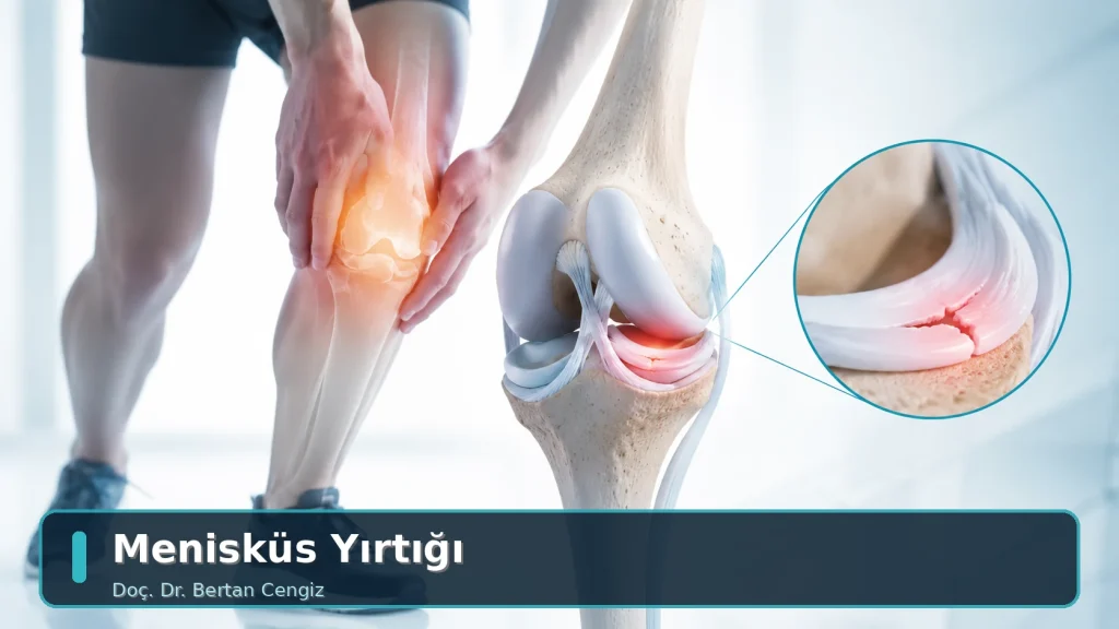
Meniscus Tear
The menisci are two «C»-shaped cartilage structures between the thigh bone and the shin bone that distribute load and absorb impact. Tears fall into two main groups: traumatic tears caused by a sudden strain, and degenerative tears that appear as the tissue wears with age. This distinction directly determines treatment.
What Does the Meniscus Do?
Each knee has an inner (medial) and an outer (lateral) meniscus. Their role is not simply to act as a cushion: by widening the contact between the joint surfaces they spread pressure over a large area, they contribute to stability during rotational movements, and they help the joint fluid nourish the cartilage. Preserving meniscal tissue therefore matters for long-term cartilage health.
The blood supply of the meniscus is not uniform. The outer zone (the red zone) is relatively well supplied and has healing potential; the inner zone (the white zone) is nourished by joint fluid and has low healing capacity. Which zone the tear lies in is the single most important factor determining whether it can be repaired with sutures.

How Does It Tear?
Traumatic tears
These are generally seen in young and active people. Typical mechanisms are the body rotating while the foot is fixed, standing up abruptly from a squat, or the knee being twisted in contact sports. They frequently accompany an anterior cruciate ligament injury.
Degenerative tears
These appear in middle age and beyond without a clear injury. The meniscal tissue gradually loses its elasticity and can tear during an everyday movement such as squatting or going down stairs. This group is often part of early-stage osteoarthritis; for that reason the treatment decision is not made on the MRI finding alone.
Symptoms
- Pain along the inner or outer joint line that can be pointed to with a finger
- Increasing symptoms on squatting, kneeling and going down stairs
- Catching, clicking or locking of the knee (an inability to straighten it fully)
- Swelling developing some hours after the injury
- A sense of insecurity during rotational movements
Locking is a particular warning: it indicates that the torn fragment is caught between the joint surfaces and calls for earlier assessment.
Diagnosis
- History: the mechanism of injury, whether there is locking or catching, and how quickly the swelling developed.
- Physical examination: joint line tenderness, meniscal tests such as McMurray and Thessaly, range of movement and assessment of ligament stability.
- X-ray: does not show the meniscus; it is requested to assess the joint space, alignment and any accompanying osteoarthritis.
- MRI: shows the site and pattern of the tear together with the cartilage and ligament structures.
Important: meniscal changes can be found on MRI in a significant proportion of people over middle age who have no symptoms at all. An imaging finding is therefore not on its own a reason to operate; symptoms, examination and function are assessed together. For more, see My MRI Report Says “Tear”.
Treatment
Non-surgical treatment first
Particularly in degenerative tears, the first approach is not surgical. Activity modification, a strengthening programme for the quadriceps and hip muscles, weight control and pain management bring marked relief for many patients. Studies report that in this group an exercise programme can give results close to those of surgery.
Arthroscopic repair (suture)
For suitable tears in the well-supplied zone the meniscus is preserved by suturing. This is preferred particularly in young patients and in tears accompanying anterior cruciate ligament surgery. The protection period after repair is longer and knee flexion may be restricted for a time.
Partial meniscectomy (limited resection)
Where the chance of a suture healing is low and the tear causes mechanical symptoms, the torn portion is removed in a limited way. The aim is to leave as much tissue as possible, because a reduction in meniscal tissue increases the load on the joint surface over the long term.
Which method is used may only become clear once the tear is seen during surgery. Always ask your physician which group you are in; your postoperative instructions depend on it. See also: Knee Arthroscopy.
After Surgery and Daily Life
Weight-bearing is generally permitted early after a partial resection; where a repair has been carried out the protection period is longer. In both cases early activation of the quadriceps is decisive in preventing muscle wasting and restricted movement. For detailed aftercare see the Knee Arthroscopy Aftercare Guide.
When Should You Seek Assessment?
- If your knee locks or does not straighten fully
- If there is catching, clicking or a sense of giving way
- If swelling developed rapidly after an injury
- If squatting and going down stairs restrict daily life
- If symptoms persist despite an exercise programme
When to Seek Medical Help Without Delay
- The knee remaining locked and unable to straighten at all
- Being unable to bear weight or take a few steps
- Obvious deformity of the knee
- Redness, warmth and severe pain in the knee together with fever
- One-sided pain, swelling and tenderness in the calf
These findings require urgent assessment. Outside office hours, go to the nearest emergency department.
Frequently Asked Questions
Does every meniscus tear require surgery?
No. Good results can be obtained with an exercise programme and pain management, particularly in age-related degenerative tears. Surgery is considered more often in tears that cause locking or mechanical symptoms, and in traumatic tears in young, active patients. The decision is based on symptoms and examination, not on the MRI.
Can a meniscus heal on its own?
Some small tears in the outer zone can heal; this is not expected for tears in the poorly supplied inner zone. However, a tear that does not heal does not always cause symptoms — the body can adapt over time.
If meniscal tissue is removed, will I develop osteoarthritis?
A reduction in meniscal tissue increases the load on the joint surface and can, over the long term, predispose to cartilage wear. That is why preserving the tissue and repairing it where possible is preferred today; where resection is needed it is kept limited.
When can I walk after surgery?
Weight-bearing is generally allowed early after a partial resection; where a meniscus repair has been carried out the protection period is longer. Your physician determines the exact timeframe according to the procedure and your healing. See our related article.
My knee makes a noise — is the meniscus torn?
Noise from the knee alone does not mean there is a tear; painless clicking is common and usually unimportant. If the noise is accompanied by pain, swelling, catching or locking, it should be assessed.
When can I return to sport?
Return is determined by criteria rather than the calendar: pain-free full movement, muscle strength close to the uninjured side, balance and the ability to perform sport-specific movements safely. This process takes longer where a repair has been performed.
Academic Work
Assoc. Prof. Bertan Cengiz, MD has scientific work published in national and international peer-reviewed journals in the field of knee arthroscopy and meniscus surgery and related areas. The publication list and verifiable academic sources are available on the Publications page.
Related Pages
- Knee Arthroscopy
- Anterior Cruciate Ligament (ACL) Injury
- Knee Osteoarthritis
- Sports Injuries
- Knee Arthroscopy Aftercare Guide
The information on this page is for general guidance and does not replace an in-person medical examination. Diagnosis and treatment require a physician’s clinical assessment. Outcomes of any treatment or surgical intervention may vary from person to person; no guarantee of a specific result or timeframe is given on this page.